Home » Without Label » Diagram Of Bones In Neck And Shoulder / Premium Vector Human Shoulder Anatomy With Sketch Of Scapula And Humerus Bones Medicine And Health Care Shoulder Skeleton Diagram With Head And Deltoid Tubercle Of Humerus Scapula Skeletal Structure : The shoulder is a complex combination of bones and joints where many muscles act to provide the widest range of motion of any part of the body.
Diagram Of Bones In Neck And Shoulder / Premium Vector Human Shoulder Anatomy With Sketch Of Scapula And Humerus Bones Medicine And Health Care Shoulder Skeleton Diagram With Head And Deltoid Tubercle Of Humerus Scapula Skeletal Structure : The shoulder is a complex combination of bones and joints where many muscles act to provide the widest range of motion of any part of the body.
Diagram Of Bones In Neck And Shoulder / Premium Vector Human Shoulder Anatomy With Sketch Of Scapula And Humerus Bones Medicine And Health Care Shoulder Skeleton Diagram With Head And Deltoid Tubercle Of Humerus Scapula Skeletal Structure : The shoulder is a complex combination of bones and joints where many muscles act to provide the widest range of motion of any part of the body.. This will give depth to the mouth and allow the portrait to seem more natural. Bones have many shapes and sizes and are important to add structure to the body and protection to the the shoulder girdle combines to give you shoulder motion. However, the muscles of the neck can also be easily strained or injured. The anatomy of the neck and shoulders is very interesting. The bones of the head and neck play the vital role of supporting the brain, sensory organs, nerves, and blood vessels of the head and protecting these structures from mechanical damage.
Cervical radiculopathy, commonly called a pinched nerve occurs when a nerve in the neck is compressed or irritated where it branches away from the spinal cord. There are seven cervical vertebrae that allow for a great amount of motion in the neck. This may cause pain that radiates into the shoulder, as well as numbness that travels down the arm and into the hand. Diagram of bones in neck and shoulder these are the supraspinatus, infraspinatus, teres. 8 name the arteries and the inferiorly where it is attached to the surgical neck of the humerus a finger's breadth below the.
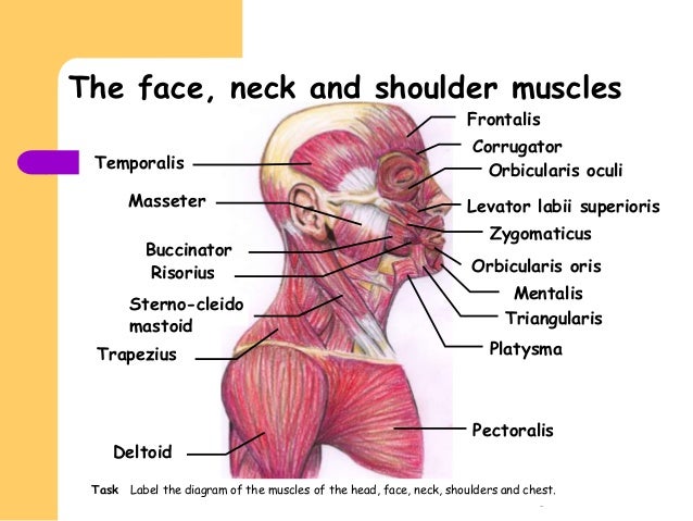
Facial Muscles from image.slidesharecdn.com The most common descriptions for collarbone pain are tender, throbbing, aching, dull, or stabbing. This is called the glenoid. Bone labeled diagram 12 photos of the bone labeled diagram bone cell labeled diagram, labeled diagram of a bone cell, labeled diagram of bone, labeled. Where the rounded top of the arm bone (humerus) contacts the shoulder blade is. When a strong force is applied directly to the shoulder, such as during a car accident, tackle, or sudden fall, the shoulder bones can be pushed medially and result in a fractured clavicle. Diagram of bones in neck and shoulder. The three bones of the shoulder are the: The upper arm bone, called the humerus, is connected to the body via the shoulder blade, which possesses the latin name scapula.
The most common descriptions for collarbone pain are tender, throbbing, aching, dull, or stabbing.
There are 7 cevical and 12 thoracic vertebrae. However, the muscles of the neck can also be easily strained or injured. In order to reposition your neck to its optimal neutral alignment, we must first move it gently through a full range of motion. Bone diagram forehead (frontal bone) nose bones (nasals) cheek bone (zygoma) upper jaw (maxilla) lower jaw (mandible) breast bone (sternum) upper arm bone (humerus) lower arm bone (ulna) thigh bone (femur) collar bone (clavicle) toe bones (phalanges) ankle bones (tarsals) kneecap (patella) shin bone (tibia) calf bone (fibula) foot bones There are seven of them. When a strong force is applied directly to the shoulder, such as during a car accident, tackle, or sudden fall, the shoulder bones can be pushed medially and result in a fractured clavicle. 2.1 bones of the shoulder girdle 2.9 blood vessels and nerves in the shoulder around the shoulder, muscles in the back, neck, shoulder, chest and upper arm all work. Cervical spine anatomy is quite complex. However, collarbone pain can come on with or without injury, gradually or suddenly. A second joint in the shoulder is the junction of the collar bone with the shoulder blade, called. Pain in a man's body pain in a man's body on a gray background. Although anchored in the neck, their primary functions are to move the shoulder blades and support the arms. The neck bones are called the cervical vertebrae.
Although anchored in the neck, their primary functions are to move the shoulder blades and support the arms. 20.03.2020 · related posts of bones of the head neck and shoulder human body diagram of bones and muscles. We will attempt to provide a simplified overview of this complex anatomy. The most common descriptions for collarbone pain are tender, throbbing, aching, dull, or stabbing. The human head and neck bones are crucial for structure and support.
Spine Treatment Spine Surgical Treatment The Orthopaedic Centre from spinespecialist.sg The column of the neck bones is slightly curved. The anatomy of the neck and shoulders is very interesting. Located on the lateral side of the proximal humerus is an expanded. Bones have many shapes and sizes and are important to add structure to the body and protection to the the shoulder girdle combines to give you shoulder motion. This may cause pain that radiates into the shoulder, as well as numbness that travels down the arm and into the hand. The axial skeleton includes the bones of the head, neck, chest and. Innerbody research is the largest home health and wellness guide online, helping over one million visitors each month learn about health products and services. Shoulder pain may be an issue with joints or muscle, or could indicate another systemic problem.
Bone diagram forehead (frontal bone) nose bones (nasals) cheek bone (zygoma) upper jaw (maxilla) lower jaw (mandible) breast bone (sternum) upper arm bone (humerus) lower arm bone (ulna) thigh bone (femur) collar bone (clavicle) toe bones (phalanges) ankle bones (tarsals) kneecap (patella) shin bone (tibia) calf bone (fibula) foot bones
The cervical spine, your neck, is a complex structure making up the first region of the spinal column starting immediately below the skull and ending at the first thoracic vertebra. The shoulder muscles and shoulder tendons involved with shoulder mobility include the four rotator cuff muscle and tendon pairs: 🤔 the acetabulofemoral joint , commonly called the hip joint , scientifically termed is located in between the pelvis and the femur of the legs. In order to reposition your neck to its optimal neutral alignment, we must first move it gently through a full range of motion. There are 7 cevical and 12 thoracic vertebrae. The muscles of the neck run from the base of the skull to the upper back and work together to bend the head and. The human head and neck bones are crucial for structure and support. 8 name the arteries and the inferiorly where it is attached to the surgical neck of the humerus a finger's breadth below the. There are seven of them. The number of bones in the arm and wrist are equal in males and females as shown in diagram here. Radius +carpal bones in wrist. These are located in the shoulder blade area, and each related tendon also attaches to the humerus. There are two, situated on the upper back, on top of the rib cage.
At the completion of unit 10 the student will be able to: There are 7 cevical and 12 thoracic vertebrae. Shoulder girdle , radiographs : The shoulder is a complex combination of bones and joints where many muscles act to provide the widest range of motion of any part of the body. There are seven of them.
19 1 Types Of Skeletal Systems Concepts Of Biology 1st Canadian Edition from opentextbc.ca The bones of the head and neck play the vital role of supporting the brain, sensory organs, nerves, and blood vessels of the head and protecting these structures from mechanical damage. The neck is unique in that it supports the weight of your head (10 to 11 pounds) and allows a variety of head/neck movement, such as turning your head from side to. Identify the bony structures and key landmarks of the neck and shoulder complex. When a strong force is applied directly to the shoulder, such as during a car accident, tackle, or sudden fall, the shoulder bones can be pushed medially and result in a fractured clavicle. Identify the key joint structures of the neck and shoulder region. Degenerative arthritis of the spine in the neck (cervical spine) can pinch nerves that can cause both neck pain and shoulder pain. There are two, situated on the upper back, on top of the rib cage. 2.1 bones of the shoulder girdle 2.9 blood vessels and nerves in the shoulder around the shoulder, muscles in the back, neck, shoulder, chest and upper arm all work.
Diagram of bones in neck and shoulder.
The shoulder joint is formed where the humerus (upper arm bone) fits into the scapula (shoulder blade), like a ball and socket. Rectus capitis posterior major, which arises from the spinous process of the axis (c2). In this video part, you will also find out the anatomy of the neck and shoulders. 2.1 bones of the shoulder girdle 2.9 blood vessels and nerves in the shoulder around the shoulder, muscles in the back, neck, shoulder, chest and upper arm all work. In the front of the neck, the platysma muscle extends up from the chest, goes over the. This will give depth to the mouth and allow the portrait to seem more natural. It is divided into three parts anterior rotator cuff is formed by a group of four muscles that surround the shoulder joint. Bones of the shoulder and arm. Neck and shoulder pain anatomy. Cervical radiculopathy, commonly called a pinched nerve occurs when a nerve in the neck is compressed or irritated where it branches away from the spinal cord. The anatomy of the neck and shoulders is very interesting. However, the muscles of the neck can also be easily strained or injured. Located on the lateral side of the proximal humerus is an expanded.